
Understanding Low-Grade Mast Cell Tumors in Dogs
When your dog is diagnosed with a low-grade mast cell tumor, it’s natural to feel overwhelmed—but there’s real reason for hope. These common skin growths in dogs often carry a favorable prognosis, especially when caught early and managed wisely. Unlike their high-grade counterparts, low-grade mast cell tumors tend to grow slowly and rarely spread aggressively. With the right veterinary guidance, many dogs go on to live full, happy lives after treatment. Understanding what this diagnosis truly means is the first step toward calm, confident decision-making.
Origins and Biology of Mast Cell Tumors
Mast cell tumors (MCTs) arise from mast cells—immune cells involved in allergic responses and wound healing. In dogs, these cells can sometimes multiply abnormally, forming tumors that vary widely in behavior. Low-grade MCTs (Grade 1) are typically well-differentiated and localized, offering a much better outlook than higher-grade forms.
- Mast Cells as Immune Players:
Mast cells normally release histamine and other chemicals during inflammation or allergic reactions—but in tumors, they replicate uncontrollably. - Why Dogs Are Prone:
Certain breeds like Boxers, Bulldogs, and Boston Terriers have higher genetic susceptibility, though any dog can develop an MCT. - Grading System Explained:
Veterinarians use grading (1 to 3) to assess how abnormal the cells look under a microscope—Grade 1 indicates low-grade, less aggressive behavior. - Location Matters:
Tumors on the limbs or trunk often behave more predictably than those on the face, groin, or mucous membranes. - Role of KIT Mutation:
Some MCTs carry a KIT gene mutation that influences growth—but low-grade tumors less commonly have this mutation, contributing to their milder nature.
While mast cell tumors sound alarming, low-grade versions are often manageable with surgery alone and rarely threaten long-term health.

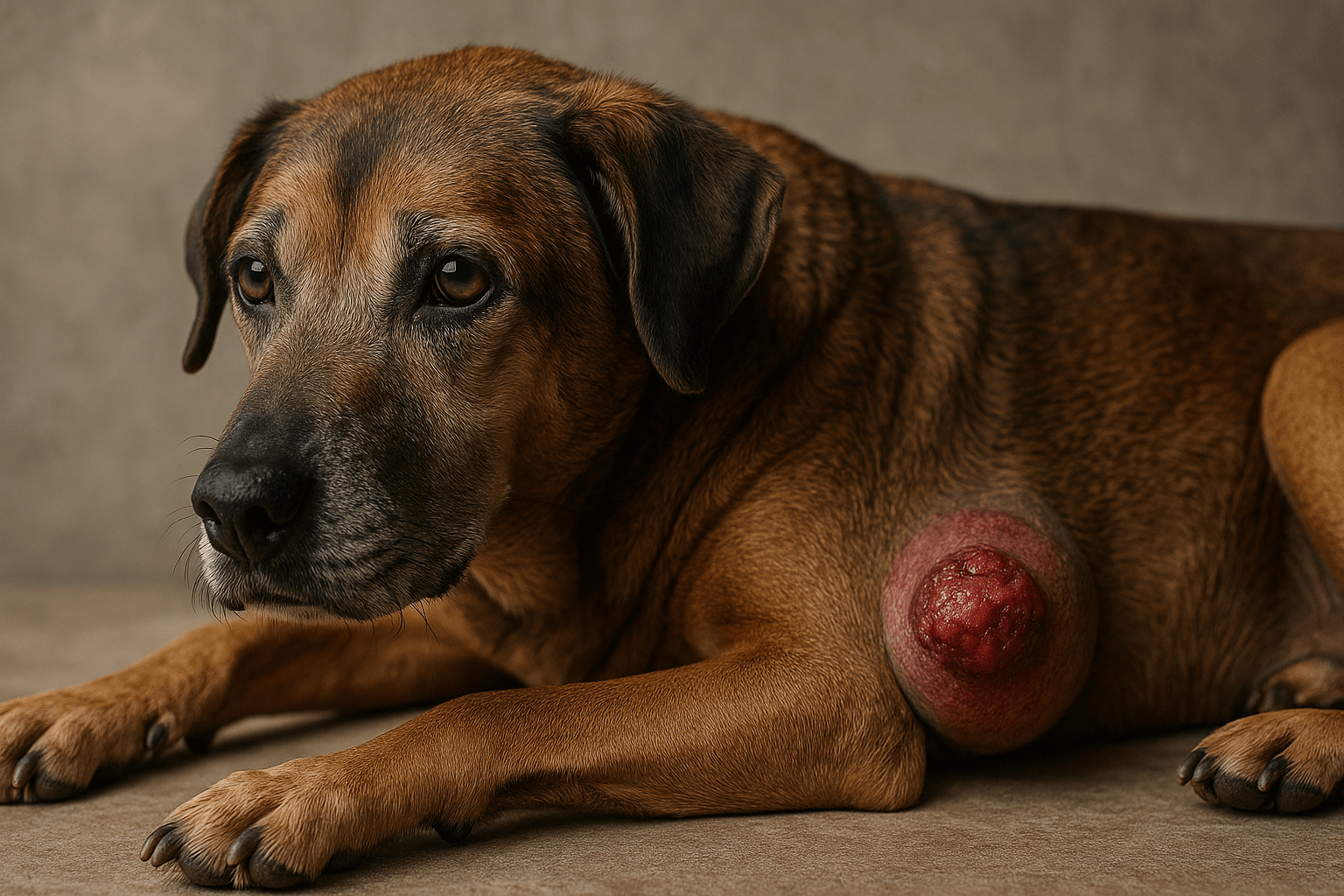
Recognizing the Signs of a Low-Grade Mast Cell Tumor
Not all skin lumps are cause for panic—but knowing what to watch for can make all the difference. Low-grade mast cell tumors often masquerade as harmless bumps, yet subtle clues can hint at their true nature. Early detection leads to simpler treatment and better outcomes.
- Variable Appearance:
These tumors may look like small, firm nodules under the skin or raised, hairless patches—sometimes red, ulcerated, or inflamed. - “Wheal-and-Flare” Response:
Gently pressing on the lump may cause it to swell temporarily due to histamine release—a classic sign of mast cell involvement. - Slow Growth Pattern:
Unlike aggressive cancers, low-grade MCTs often grow gradually over weeks or months, not days. - Isolated Lesion:
Most low-grade tumors appear as a single lump, not multiple growths—a reassuring indicator of localized disease. - Minimal Systemic Symptoms:
Dogs usually feel well, with no vomiting, lethargy, or appetite loss unless the tumor is irritated or infected.
Because these signs can mimic benign cysts or insect bites, any new or changing skin mass should be evaluated by a vet.
Check this guide 👉Types of Dog Tumors: Best 7 Expert Tips!
Check this guide 👉Vascular Tumors in Dogs: Best 7 Expert Tips!
Check this guide 👉Dog Brain Tumor Symptoms: Best 7 Expert Tips!
| Favorable Indicators | Monitoring & Care Needs |
|---|---|
| Single, small tumor (<3 cm) | Monthly skin checks for new lumps |
| Located on trunk or limb | Post-surgery wound care for 10–14 days |
| No lymph node involvement | Histopathology report review with vet |
| Grade 1 on biopsy | Antihistamine support if recommended |
| Clear surgical margins achieved | 6-month recheck ultrasound (if advised) |
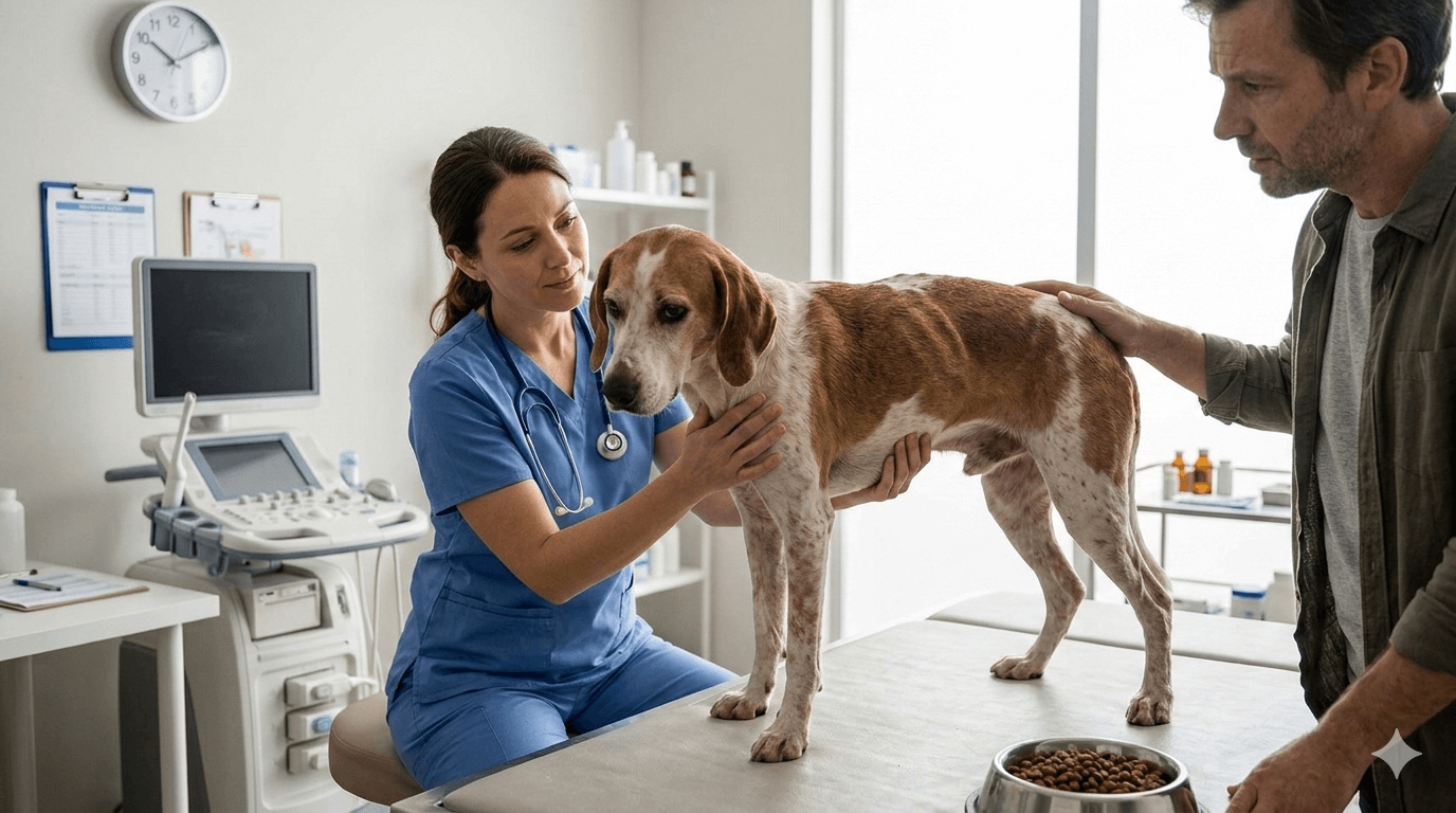
Diagnosis and Staging Process
Confirming a low-grade mast cell tumor involves more than just a visual exam—it requires precise diagnostic steps to rule out hidden spread and ensure accurate grading. Your vet will likely recommend a series of tests to gather a complete picture before deciding on treatment.
- Fine Needle Aspirate (FNA):
A quick, minimally invasive test where cells are extracted with a thin needle—often the first clue that a lump is a mast cell tumor. - Surgical Biopsy:
Definitive grading requires removing the entire mass (or a core sample) for histopathology—a pathologist examines cell structure under a microscope. - Lymph Node Assessment:
Local lymph nodes may be aspirated or imaged to check for microscopic spread, even if they appear normal. - Abdominal Ultrasound:
Recommended in some cases to evaluate the spleen, liver, and internal organs—though low-grade tumors rarely involve these areas. - Bloodwork and Urinalysis:
These help assess overall health and detect elevated mast cell byproducts like tryptase or histamine metabolites.
Together, these steps ensure your dog’s tumor is correctly classified—and that the treatment plan matches its true risk level.
Treatment Options for Low-Grade MCTs
The good news? Most low-grade mast cell tumors are cured with complete surgical removal alone. Unlike high-grade cases, chemotherapy or radiation is rarely needed—making treatment straightforward and less stressful for both pet and owner.
- Wide-Excision Surgery:
Veterinarians remove the tumor plus a margin of healthy tissue (usually 1–2 cm) to ensure no cancer cells are left behind. - Pathology Confirmation:
The removed tissue is sent to a lab to confirm clean margins—if margins are narrow, a second surgery may be advised. - No Additional Therapy Typically Needed:
If the tumor is truly Grade 1 with clear margins, no further treatment is usually required—just monitoring. - Antihistamines for Symptom Control:
Some vets prescribe Benadryl or similar to reduce histamine-related inflammation before or after surgery. - Pain Management and Recovery:
Post-op pain control, restricted activity, and an E-collar help ensure smooth healing with minimal complications.
With proper surgical care, many dogs never experience recurrence—giving you peace of mind and your pet a return to normal life.
Prognosis and Long-Term Outlook
A low-grade mast cell tumor diagnosis is one of the more optimistic scenarios in canine oncology. Most dogs enjoy excellent long-term survival with no further issues after successful surgery—especially when the tumor is caught early.
- High Cure Rate:
Studies show over 90% of dogs with completely excised Grade 1 MCTs remain tumor-free for life. - Low Recurrence Risk:
If surgical margins are clean, recurrence at the same site is uncommon—typically under 5%. - Rare Metastasis:
Spread to lymph nodes or organs is exceptionally rare with true low-grade tumors. - Normal Life Expectancy:
These dogs usually live out their natural lifespan with no cancer-related limitations. - Importance of Follow-Up:
Even with a great prognosis, regular vet check-ups help catch any new skin masses early—because dogs can develop multiple MCTs over time.
In short, a low-grade diagnosis is often the beginning of the end of the problem—not the start of a long battle.
Potential Complications and Red Flags
While low-grade mast cell tumors are generally benign in behavior, vigilance is still key. A small percentage may behave unpredictably, or a misgraded tumor could pose hidden risks. Knowing warning signs helps you act fast if needed.
- Incomplete Surgical Margins:
If pathology shows tumor cells at the edge of the removed tissue, recurrence risk rises significantly. - Unexpected Aggression:
Rarely, a tumor labeled “low-grade” may act more aggressively—highlighting the need for expert pathology review. - Multiple New Tumors:
Some dogs are genetically prone to developing new MCTs elsewhere—prompt evaluation of any new lump is essential. - Systemic Mastocytosis:
Extremely rare in low-grade cases, but signs like vomiting, diarrhea, or low blood pressure warrant immediate vet attention. - Infection or Ulceration:
An irritated or open tumor can cause pain, bleeding, or secondary infection—requiring medical management before surgery.
Staying alert doesn’t mean living in fear—it means empowering yourself to protect your dog’s health proactively.
Prevention and Daily Care Strategies
Though you can’t always prevent mast cell tumors, smart daily habits and routine monitoring can catch issues early and support your dog’s immune resilience—especially for at-risk breeds.
- Monthly Skin Checks:
Run your hands over your dog’s body once a month, noting any new bumps, especially on the trunk, limbs, or head. - Sun Protection for Light-Coated Dogs:
While not directly linked to MCTs, UV exposure can damage skin—use pet-safe sunscreen on sensitive areas if needed. - Balanced, Anti-Inflammatory Diet:
Omega-3 fatty acids, antioxidants, and high-quality protein support immune function and skin health. - Avoid Chronic Skin Irritation:
Repeated trauma or inflammation may theoretically influence tumor development—treat allergies and skin infections promptly. - Genetic Awareness:
If you own a high-risk breed, discuss early screening options with your vet—even before a lump appears.
Preventive care isn’t about eliminating risk entirely—it’s about stacking the odds in your dog’s favor every single day.
Frequently Asked Questions
Can a low-grade mast cell tumor become high-grade over time?
True low-grade (Grade 1) tumors rarely transform, but new, unrelated high-grade tumors can develop—underscoring the need for ongoing monitoring.
Is my dog in pain from a low-grade MCT?
Usually not—unless the tumor is ulcerated, infected, or located in a sensitive area. Most dogs show no discomfort.
How quickly should surgery happen after diagnosis?
Ideally within 2–4 weeks. These tumors grow slowly, but early removal reduces inflammation and simplifies healing.
Do I need an oncologist for a low-grade MCT?
Not always—many general vets handle complete excision successfully. However, an oncologist can help if margins are unclear or if you want expert confirmation.
Will my dog need chemotherapy?
Almost never for confirmed low-grade, completely excised tumors. Chemotherapy is reserved for high-grade, incomplete, or metastatic cases.
A Diagnosis That’s Manageable—Not Miserable
Hearing “mast cell tumor” can send your heart racing—but with a low-grade diagnosis, the path forward is often clear, calm, and highly successful. These tumors, while concerning at first glance, respond beautifully to timely, straightforward care. Your love, attention, and partnership with your vet are the most powerful tools in your dog’s healing journey.
With surgery and smart follow-up, your pup can return to zoomies, naps in the sun, and tail-wagging routines without cancer holding them back. Trust the process, lean on your vet team, and remember: low-grade doesn’t mean “no hope”—it means “excellent chance.” And that’s a story worth celebrating, one healthy day at a time.

Is Rawhide Bad for Cats? Best 7 Expert Tips! – Discover the risks, safe alternatives, and expert advice to keep your feline friend healthy and happy.

Is Rawhide Bad for Dogs? Best 7 Expert Tips! – Discover the risks, benefits, and safer alternatives to rawhides for your dog’s chewing needs.

Understanding Anorexia in Cats: Best 7 Expert Tips! – Learn why cats stop eating, spot warning signs, and discover how to help your feline regain appetite safely.
Understanding Anorexia in Dogs: Best 7 Expert Tips! – Learn causes, symptoms, and solutions to help your dog regain appetite and stay healthy.